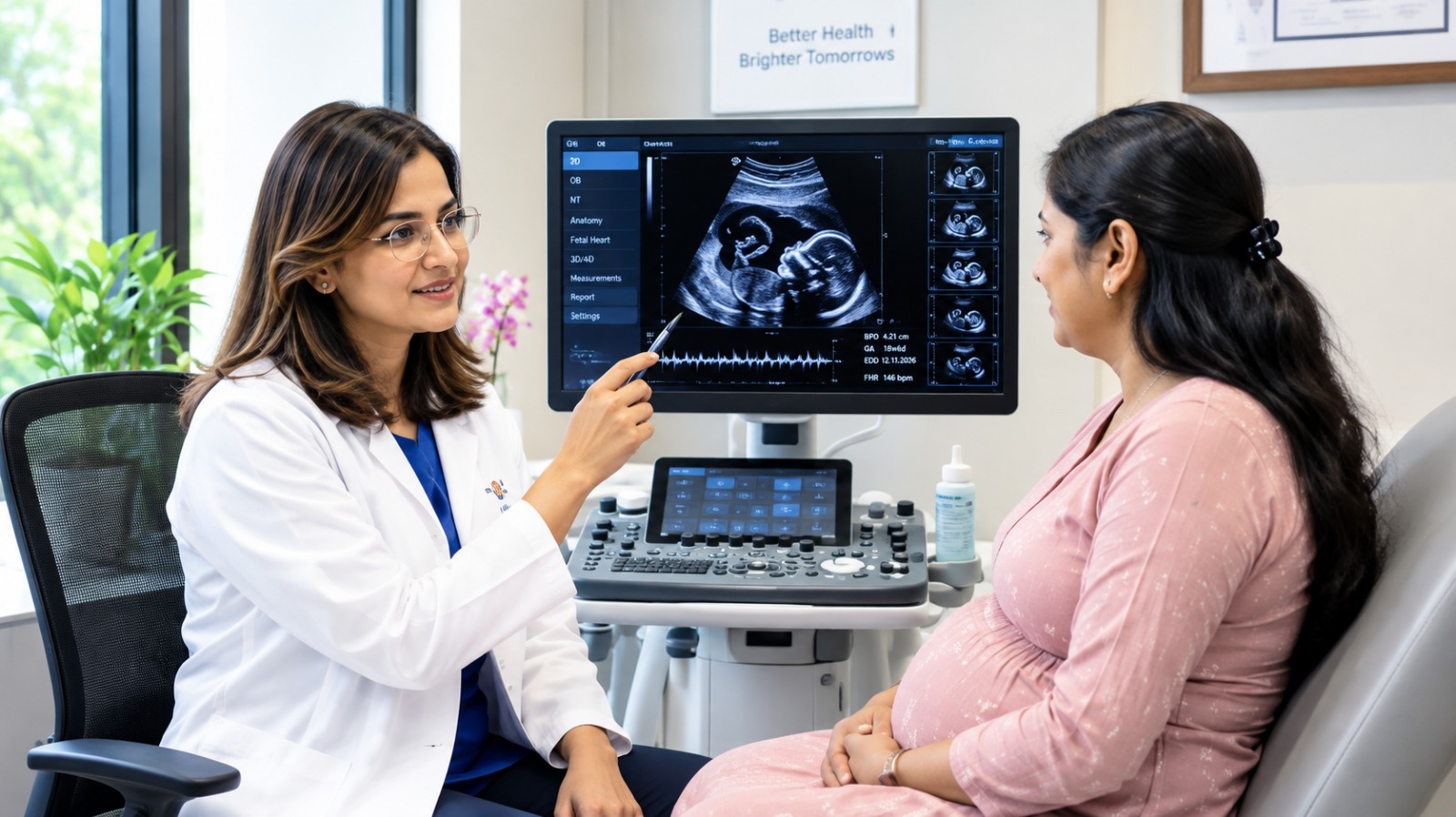
Fetal Echocardiography: Why Your Baby’s Tiny Heart May Need a Closer Look
 161 Reads
 4 minutes read

“Doctor, my Level 2 scan was normal. Then why do I need a fetal echocardiography?”
This is one of the most common questions I hear from expectant parents. And I understand why. Being asked to undergo another scan can immediately make parents worry that something may be wrong with their baby.
But a fetal echocardiography does not necessarily mean that a heart problem has been found. Often, it simply means that your doctor wants to take a closer look at one of the most important organs your baby is developing.
The Level 2 scan looks at the baby. The fetal echo looks closely at the heart.
A Level 2 or anomaly scan examines your baby’s overall development, including the brain, face, spine, limbs, abdomen, placenta and other major structures.
A fetal echocardiography is a specialised ultrasound examination focused specifically on the baby’s heart.
By around 18–24 weeks, the heart is still tiny, but it is already working continuously. During the examination, we carefully look at the heart chambers, valves, major blood vessels, heartbeat and blood flow, as well as signs of possible structural abnormalities.
Think of the anomaly scan as looking at the whole picture, while a fetal echo takes a much closer look at one particularly important part.
Why might your doctor recommend it?
A fetal echo may be advised when there is a particular reason to examine the baby’s heart more closely. These may include:
- A possible heart abnormality seen during the anomaly scan
- An increased NT measurement
- A family history of congenital heart disease
- Diabetes in the mother
- IVF pregnancy
- Twin or multiple pregnancy
- An unusually fast, slow or irregular fetal heartbeat
- Certain maternal medications or infections
- Difficulty getting a complete view of the heart during the routine scan
Importantly, being referred for a fetal echo does not mean that your baby definitely has a heart defect.
Can it detect every heart problem?
No medical test can detect every condition before birth.
However, fetal echocardiography provides a detailed assessment of the baby’s heart and can identify many important congenital heart problems during pregnancy. A normal examination is reassuring, but it does not replace routine pregnancy care or later assessments when they are advised.
Early information can also be valuable. If a significant heart condition is detected, parents and doctors can discuss further monitoring, specialist consultations and plans for delivery and care after birth.
What happens during the scan?
The examination is simple and non-invasive. A probe is gently moved over the abdomen using ultrasound gel. There are no injections or X-rays involved.
Sometimes the baby is in a position that makes the heart difficult to see clearly. In that situation, you may be asked to change position, walk around for a few minutes or return for another look.
I often tell parents, “Your baby’s first game of hide-and-seek may already have begun.”
One thing I want every parent to remember
A fetal echocardiography is not a scan designed to create fear. It is a scan designed to replace uncertainty with better information.
The Level 2 scan looks at the whole baby. The fetal echo looks closely at the heart. One complements the other; neither replaces the other.
As parents, you may see a tiny heart beating on the screen. As a radiologist, I see chambers, valves, blood flow and developing structures. But we are both looking at the same thing: the beginning of a lifetime.
At Pacific OneHealth, the focus is not simply on performing a scan, but on helping families understand what the images mean and what they should do next.
When it comes to your baby’s heart, it is always better to know than to wonder.
(The author is Principal Consultant – Radiology, Pacific OneHealth Hospital, Lajpat Nagar, New Delhi, Specialising in USG-Guided MSK Interventionist Radiology.)